Strong signals for improved diagnostics
For the first time, scientists have succeeded in in amplifying the nuclear spin signal of a DNA biomolecule, which specifically binds to cancer cells, by several orders of magnitude.
09/06/2023 · News · DWI - Leibniz-Institut für Interaktive Materialien · Mathematik, Natur- und Ingenieurwissenschaften · Forschungsergebnis
Scientists at the DWI - Leibniz Institute for Interactive Materials and the Institute of Technical and Macromolecular Chemistry at RWTH Aachen University have succeeded for the first time in amplifying the nuclear spin signal of a DNA biomolecule, which specifically binds to cancer cells, by several orders of magnitude. The signal amplification methodology used is called para-hydrogen induced hyperpolarization (PHIP). The results pave the way for highly sensitive molecular magnetic resonance imaging with targeted contrast agents and have the potential to significantly expand the fields of application in medical diagnostics.
Combination of DNA and PHIP
A research team from the DWI - Leibniz Institute for Interactive Materials and the Institute of Technical and Macromolecular Chemistry at RWTH Aachen University has used a creative approach to show that there is more potential in the application of PHIP then currently used: They have succeeded for the very first time in equipping a targeted DNA molecule from the laboratory, which specifically targets cancer cells, with a hyperpolarizable marker and amplifying its magnetic resonance signal by several orders of magnitude. Here, para-hydrogen served as the hyperpolarization source bound to the marker. Never before has DNA been hyperpolarized, which is why this work , led by Prof. Dr. Andreas Herrmann, Prof. Dr. Jürgen Klankermayer and Dr. Meike Emondts, lays the foundation for further research in this area. These modified DNA molecules show good potential for use as contrast agents in molecular imaging: on the one hand, the DNA molecules can be selected individually to target specific cells or pathogens, and on the other hand, the significant signal amplification solves the problem of the usually low signal intensity of molecular magnetic resonance imaging (MRI). For this new method to be considered for clinical applications in the future, the lifetime of hyperpolarization needs to be extended and an efficient purification method of DNA after reaction with para-hydrogen needs to be developed. These are aspirations of ongoing and future research.
The team's long-term goal is to make the technology presented here applicable in medical diagnostics through further research to enable highly sensitive molecular MRI. The ability to significantly amplify MRI signals opens up the potential to detect abnormalities at the cellular level even earlier and thus improve treatability. Since the signal intensity in the new method is independent of the magnetic field strength, the use of portable, low-cost MRI scanners for molecular imaging could be envisioned in the future. This would significantly improve applicability and open up new possibilities, such as molecular MRI during surgery.
Significance of imaging techniques in clinical application
Medical imaging techniques allow physicians to visualize the internal state of the body and detect potential abnormalities or diseases. For example, by using imaging technologies such as X-ray, computed tomography (CT), MRI or ultrasound, they can identify tumors, fractures, infections, injuries and other health problems. In addition, when preparing for upcoming treatments or monitoring potential changes in the affected tissue, these technologies can help create a treatment plan tailored to the patient and plan interventions more accurately. Ultimately, imaging techniques also play a crucial role in the early detection of diseases.
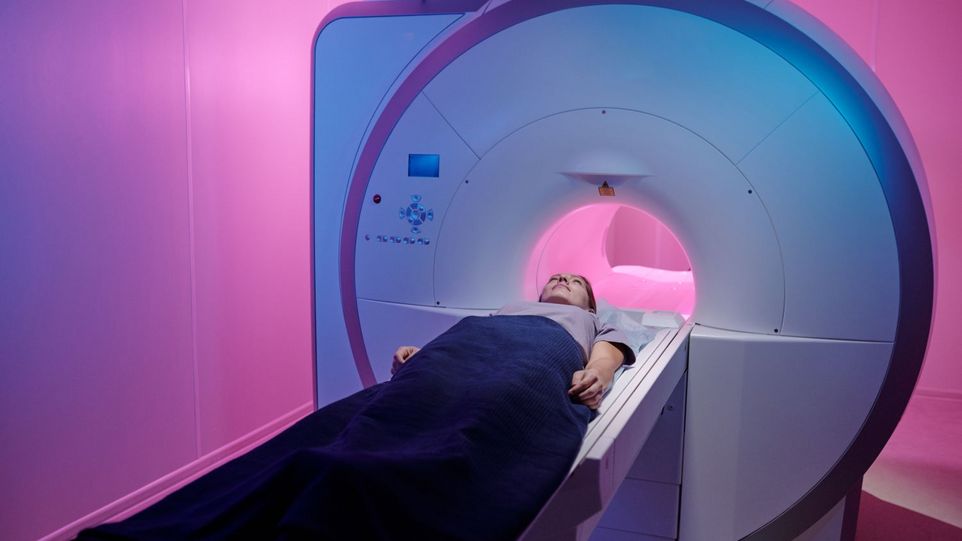
Magnetic resonance imaging and hyperpolarization
MRI is one of the imaging procedures that has become an indispensable part of everyday clinical practice in medical diagnostics. An external magnetic field initially aligns the nuclear spins. Radio waves are used to resonantly excite the nuclear spins. This results in a signal which is measured and from which an image is derived. The stronger the external magnetic field, the better the MRI signal. In order to receive the best possible signals and thus obtain good MRI images, very strong magnetic fields are therefore usually used (about 20,000 to over 50,000 times stronger than the earth's magnetic field).
By means of so-called hyperpolarization, MRI signals can be significantly amplified independently of the applied magnetic field. The alignment of the nuclear spins is then not achieved by applying an external magnetic field, but by interactions with a molecular hyperpolarization source, such as para-hydrogen. In this way, even low-threshold signals can be displayed that are difficult or impossible to detect without hyperpolarization.
Molecular imaging
Molecular imaging allows the imaging of physiological processes at the molecular level in the human body, such as changes in cellular metabolism or in the structure and function of the cell. In many diseases, such cellular processes are disturbed: for example, in some cancers, certain proteins are increasingly produced on the cell surface. Such processes can then be detected by using specific contrast agents. These consist of a targeting unit capable of specifically addressing biomarker targets in the body - such as the aforementioned cell surface proteins - and a signaling molecule that can be detected by the selected imaging modality. This enables both the localization and identification of degenerated cells, as well as other pathogens in the body. MRI represents an excellent imaging modality in this regard due to its high spatial resolution and non-invasive nature. Molecular MRI thus holds enormous potential for the early detection, diagnosis and treatment monitoring of tumors, for example, but is still in its early stages from a clinical perspective. Often, one of the limiting factors for imaging molecular processes is sensitivity. Hyperpolarization can be used to enhance signals in molecular MRI, but this has only been demonstrated for small metabolites. However, with the technique introduced in this work, hyperpolarization of larger molecules is also possible.
Further information and contact
Presse release - DWI - Leibniz-Institut für Interaktive Materialien